You may be asking “what is a ‘visual field‘ and how on earth do you test it?” Even if you weren’t, we’re going to tell you anyway. Join us for this article about the different machines we use in our office.
If you’ve already done this test, you may think it’s more of a torture device than a high-tech piece of ophthalmology equipment. So we hope to demystify the machine and give you a better understanding of what we’re doing.
What is a Visual Field?

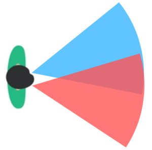
Your visual field is the range that you can see. It’s basically a cone that projects from each eye; the maximum you can see to the left, right, top, and bottom of each eye.
When we perform visual field testing, we make a map of this field. It’s a bit like a topography map and shows places you can see light better than others.
Because your two eyes overlap (as shown in the picture here), you may have blind spots in your vision that you didn’t know about. In fact, everybody has a “physiologic” blind spot. You can check it yourself by holding both of your thumbs up next to each other in front of you. Close your left eye and look at your left thumb. Move your right thumb out to the right slowly and keep looking at your left thumb. You’ll notice the tip of your right thumb disappear, because that’s your blind spot!
Blind spots don’t always look like black spots in your vision. That’s because your brain is very good at filling in what you’re seeing.
Why Does Your Doctor Order Visual Field Testing?

Your visual acuity (how sharply you can see) and your visual field are the two most important tests of your eyes function. When you come in for a comprehensive eye exam, we do a “gross” (or low-sensitivity) test of your visual field, by checking if you can see our fingers in the periphery when you look straight ahead.

If that testing shows something abnormal, your doctor will likely order a more detailed visual field test. Sometimes various eye conditions or medications can impact your visual field as well. Glaucoma is the most common reason we perform visual field testing. It’s a progressive disease that degrades vision in specific patterns.
Visual field testing isn’t only about your eyes, though. Because your eyes don’t really “see.” They collect light information, convert it to nerve impulses, and send that to your brain. Your brain figures out what you’re seeing. If the nerve signal is interrupted between your eyes and the back of your brain (your occipital lobe is where vision is processed), that can cause blind-spots as well.
Detecting strokes and tumors from visual field testing allows us to get you the help you need faster.
What is Visual Field Testing Like?
Unfortunately, testing your visual field can be very boring. Your technician will escort you to the visual field machine, which has 2 sides to the headrest and a lens to help you focus inside the testing area. We’ll calculate the best lens to use based on the glasses prescription of each of your eyes. Because the fields of your two eyes overlap, it’s important to put a patch over one eye during testing.
Your technician will adjust the machine up or down for your comfort and will move you inside the headrest to keep your eye centered. They’ll review the instructions for the test, something like this:
- There will soon be little flashes of white light in random locations inside the machine. Please press your response button every time you see one of the flashes.
- Keep your chin resting down and your forehead pressed forward in the head rest.
- Blink normally.
- Keep focused on the yellow/orange light the entire time, please do not look around.
The testing takes somewhere between 3 and 10 minutes for each eye, depending on the specific testing strategy your doctor orders. While this is an incredibly long time to stay focused, the technology has improved dramatically and used to be a 15-20 minute test per eye.
What Do Visual Field Test Results Look Like?

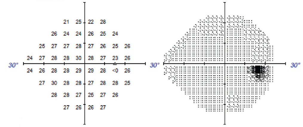
Here we see an example of the map generated by our automated visual field testing machine. On the right is a grayscale picture of a normal eye, with that physiologic blind spot we all have. On the left, is the raw data of the test.
Our visual field tests are “threshold” tests, meaning they find out the dimmest light you can see in any particular spot. Dimmer lights are more attenuated. Decibels are the units of measured attenuation (named after telephone inventor, Alexander Graham Bell). The brightest light stimulus the machine can show has been attenuated by 0 decibels. The absolute dimmest light it can show has been attenuated by about 40 decibels. So on the raw data, the higher numbers mean better vision in that spot. If you look at the numbers where the black spot is on the grayscale picture, you’ll see it says <0, meaning no light was seen there even with the brightest light available.
Summary
Visual field testing is an important part of checking your visual function. Your doctor orders this testing based on different signs or symptoms of vision loss and for specific disease monitoring. It can be tedious and it’s common to feel like you didn’t get it right.
We hope this article has been helpful in helping you understand what visual field testing is, why we do it, and what it shows your doctor. Please join us next time for the next article in our series looking into the different equipment we use to examine your eyes!
